Image: medgurus.org
A neurosurgeon in Lafayette, Louisiana, and a graduate of Rush Medical College in Chicago, Dr. Ilyas Munshi has owned a private practice providing neurological diagnosis and surgery since 2001. Dr. Ilyas Munshi devotes his time to brain, spinal, and peripheral nerve conditions, and is well-versed in computer-assisted, image-guided surgery, also called neuronavigation.
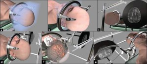
Image-guided neuronavigation helps doctors to plan the safest and most efficient approach for a surgical procedure by allowing the surgeon to view the internal area on which he or she is about to operate.
Neuronavigation uses the principle of stereotaxis: generic points, present in every brain, are combined with MRI or CT scan images of the patient’s cranium, offering a personalized view of the brain in question. This technology lets the doctor try out different points of access – to reach a tumor, for instance – before making even a single cut.
Cranial surgery benefits substantially from neuronavigation technology. The ability to plan ahead means the opening that must be made in the skull can be smaller and better placed, for optimal access. Tumors located in important areas of the brain can be removed with considerably less risk of damage to the patient’s motor and mental functions.